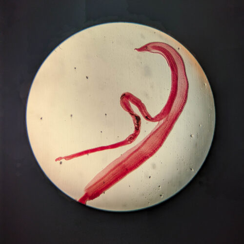
A routine medical examination in the Philippines uncovers a rare case of neuroschistosomiasis after a patient presents with sudden seizures. Medical experts utilized a vintage 1950s laboratory test that confirmed the diagnosis when reference eggs visibly grew antibody tails.
Sudden Neurological Onset Sparks Emergency Response
Out of nowhere, a woman's right arm and leg started shaking uncontrollably. In the moments leading up to the experience, she seemed well; she had no headache, confusion, or weakness. But for about a minute, half of her body juddered. The patient, a rice farmer residing in the Philippines, immediately sought medical attention and headed to the emergency department for professional evaluation.
Upon her arrival, attending medical staff administered a comprehensive physical exam and initially found nothing out of the ordinary. However, looking deeper into her condition required advanced diagnostic imaging of her cranial structure. When physicians took a look at her brain using magnetic resonance imaging (MRI), they revealed a striking bright cluster of nodules surrounded by fluid located specifically in her left frontal lobe.
Faced with these unusual imaging results, the clinical team formulated an immediate hypothesis regarding the origin of the nodules. Based on the geographic region and the visual presentation of the brain scan, the doctors suspected that the foreign bodies were worm eggs. This prompted an extensive investigation into an uncommon and complex parasitic complication that would soon require specialized diagnostic techniques.
Investigating the Complex Life Cycle of Blood Flukes
In a detailed case report published in the latest issue of the New England Journal of Medicine, doctors write that they suspected the woman had neuroschistosomiasis, an uncommon complication of an infection with a Schistosoma parasitic worm, commonly known as a blood fluke. In the Philippines, the most prevalent and concerning fluke species is Schistosoma japonicum. This specific parasite is also endemic to China and parts of Indonesia; despite its misleading geographical nomenclature, it has been successfully eliminated from Japan.
S. japonicum, much like its close biological relatives, possesses a diabolical life cycle. The cycle starts with its eggs being shed in the urine or feces of various infected animal hosts, including humans. When environmental conditions are optimal, the eggs hatch into a free-swimming larval form. These microscopic larvae actively seek out specific types of aquatic snails, which they bore into and inhabit to go through two distinct life stages before continuing their journey.
Late-stage larvae then burst out of the intermediate snail hosts, grow a forked tail, and swim vigorously to find a definitive animal host. These tailed larvae, known scientifically as cercariae, possess the remarkable and dangerous ability to tunnel directly through intact skin. Once inside, they take a complex trip through the vascular system - visiting the lungs, the heart, and finally settling in the liver, where they develop into fully mature adults.
Chronic Infection and Central Nervous System Complications
The adult male and female worms live paired together in a permanent union, with the slender female worm tucked neatly into a specialized gynecophoral groove within the thicker male. Adult worms typically migrate to large blood vessels in the abdomen that connect the liver directly to the intestinal tract. From this vascular hub, the paired couples continuously spew out microscopic eggs that make their way into feces and urine to perpetuate the species.
Remarkably, adult worms often go entirely undetected within blood vessels, producing virtually no noticeable clinical symptoms, which allows the parasitic infection to become chronic over extended periods. It is typically the microscopic eggs that cause pathological problems and ultimately give the worms' presence away. These deposited eggs can spur severe localized inflammation, tissue fibrosis, and complex organ-specific complications throughout the body.
On rare and unpredictable occasions, S. japonicum eggs manage to reach the central nervous system, creating severe medical complications. It is not entirely clear how this aberrant migration happens; medical experts speculate that it may occur when either the adult worms or their eggs get diverted into an unusual anatomical place, possibly due to a heavy parasitic infestation or underlying vascular damage within the host.
Vintage Laboratory Testing Confirms the Diagnosis
However the aberrant migration happens, when parasite eggs get lodged directly into the brain tissue, they are frequently linked to sudden seizures or specific neurological deficits. The realistic possibility of S. japonicum eggs residing within the woman's brain seemed to fully explain her abrupt seizure episode, prompting her medical team to initiate a rigorous battery of diagnostic tests to confirm the clinical suspicion.
Because there is no single definitive go-to test for schistosomiasis, clinicians routinely perform various complementary tests to accurately confirm an active infection. The woman's medical team first turned to an old-school laboratory test called the circumoval precipitin test, which was originally developed back in the 1950s. In this specialized procedure, doctors obtain a reference sample of S. japonicum eggs and flood them with the patient's blood serum.
If the patient is suffering from an active infection, their serum will contain specialized antibodies capable of targeting the parasitic egg. In this immunological event, the antibodies can grab onto specific molecules dangling off the outside of the egg as well as molecules actively secreted by the egg. In the laboratory test, this specific biological reaction leads to a visible, tail-like structure of bound antibodies forming on the outside of the reference egg.
Definitive Results and Clinical Implications
While medical professionals note that this vintage test does not always yield positive results in every clinical scenario, in this particular patient's case, it produced a remarkably clear and unmistakable outcome. The test eggs grew weird, distinct tails in the lab test, definitively indicating an active S. japonicum infection. There is no single definitive test for these worms, but the tail is fairly convincing.
To solidify the medical evaluation further, another diagnostic test independently found a high concentration of S. japonicum eggs in the woman's stool sample. Specifically, laboratory analysts counted a staggering 190 eggs per gram of feces, providing overwhelming quantitative evidence of the systemic parasitic burden.
Combined with the initial MRI brain scan results suggesting the presence of foreign egg clusters in her left frontal lobe, the multidisciplinary clinical team successfully diagnosed her with cerebral schistosomiasis. This comprehensive diagnostic triumph highlights the enduring utility of historical laboratory methods in solving complex modern medical mysteries.
Reporting synthesized under Nexvoro.tech Editorial Standards • Referenced via Ars Technica
Verified Dispatch